Laser & microscope
Microscope dentistryin Cape Town
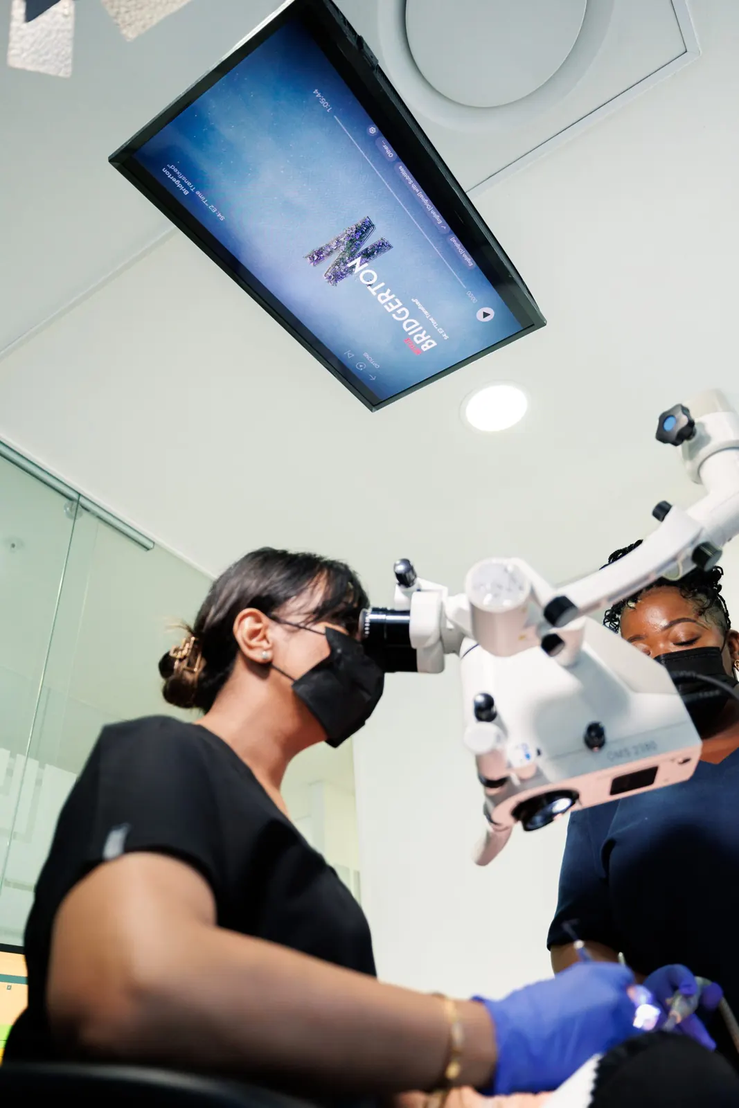
Microscope dentistry at Singh Advanced Dentistry in Gardens, Cape Town uses a dental operating microscope to magnify and light the treatment area during root canals, fillings and other detailed work.
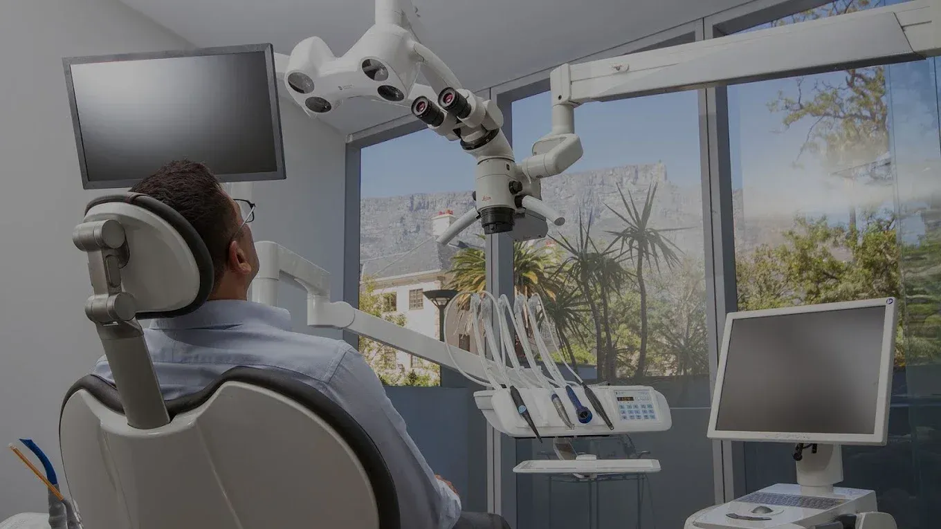
Microscope dentistry, sometimes called magnified dentistry, uses a Leica dental operating microscope so the dentist can see fine detail while planning and carrying out treatment.
At a glance
- Who it's for
- Root canal, restorative and precision work
- Time in the chair
- —
- Number of visits
- —
- Recovery
- —
What the microscope changes
Teeth are small, and many of the details that matter are smaller still. The microscope combines high magnification with focused light, so the dentist can see fine detail that the naked eye can miss.
That includes hairline cracks, the openings of root canals, and the edges where fillings and crowns meet the tooth.
- Root canal treatment
- Fillings and inlays
- Crown fitting
- Checking old restorations
Where we use it
We use the microscope for root canal treatment, fillings and inlays, fitting crowns, and checking older fillings and crowns for wear or gaps.
Your dentist will explain at your consultation whether it is useful for your treatment.
Same chair, same appointment, with a closer view of the detail.
What to expect
Treatment happens in the same chair as any other appointment. You may notice the microscope arm positioned above you while the dentist works.
Because of the care taken, treatment can take a little longer than you might expect. We'll tell you how long to plan for.
How it works
Step by step.
- 01
Consultation
We examine your teeth and explain whether magnification will help with your treatment.
- 02
Your plan
You receive a clear treatment plan and costs before anything begins.
- 03
Treatment
The dentist works through the microscope, at a pace that suits you.
Your journey
Microscope dentistry, step by step.
- Step 1
Consultation
We examine your teeth, talk through your goals and explain the options and costs.
- Time
- 45–60 min
- Visits
- 1 visit
- Step 2
Magnified treatment
Your treatment is carried out through the dental operating microscope, so fine detail can be seen clearly.
- Visits
- Depends on treatment
Practice to confirm
Times are typical and vary from person to person. We'll confirm yours at your consultation.
FAQs
Questions answered.
Is it uncomfortable?
The microscope doesn't touch you. It sits above the chair on an arm, and treatment is otherwise the same as a normal appointment, with local anaesthetic where needed.
Does every treatment use it?
No. We use it where fine detail matters, such as root canal treatment, fillings, inlays and crown fitting. Your dentist will tell you if it will be used.
How is it different from loupes?
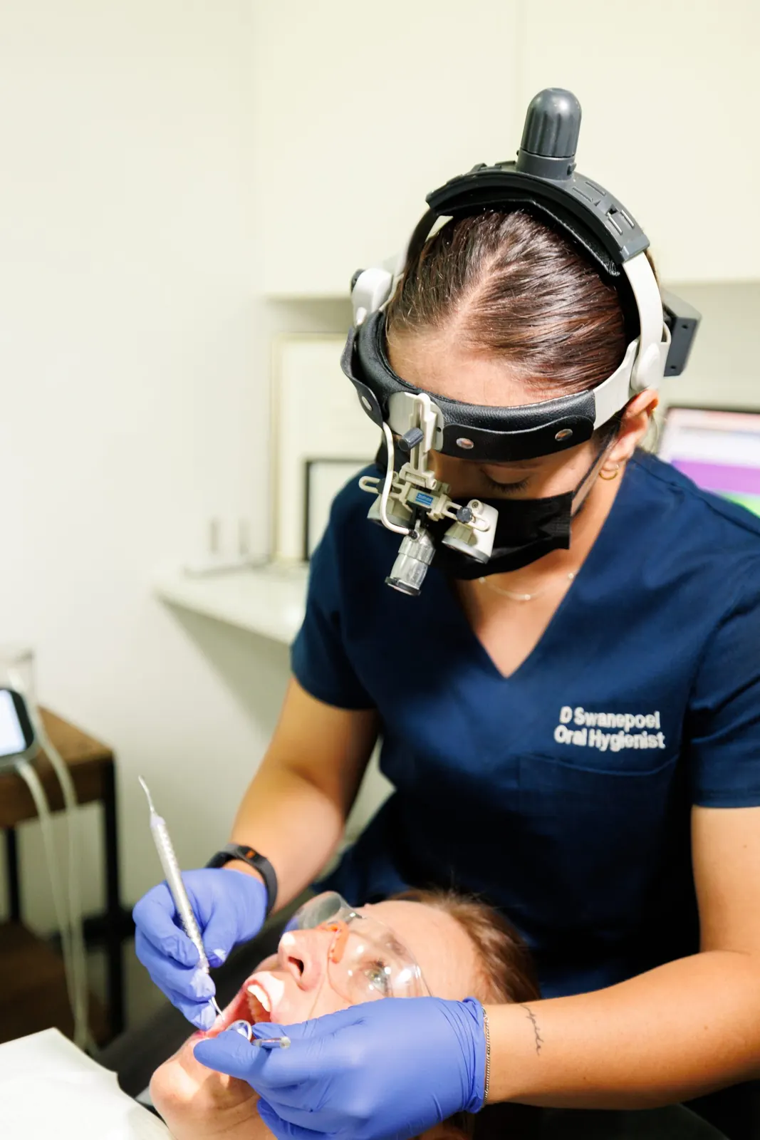
Loupes are magnifying lenses worn like glasses. A dental operating microscope can magnify further and has its own built-in light, which helps when looking at very small areas.
Related treatments